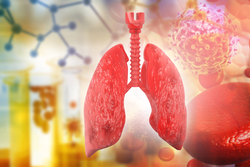
Cells Grown in Artificial Organs May Be Most Viable for Lung Disease Research


Growing cells in artificial tracheas or lungs is a better approach to model native organs than cells cultured in spheroids or in a plastic filter, according to a study, suggesting that these platforms may be more suitable to study cell-based therapies for lung diseases such as cystic fibrosis (CF).
The study, “Platform Effects on Regeneration by Pulmonary Basal Cells as Evaluated by Single-Cell RNA Sequencing,” was published in the journal Cell Reports.
The airways and lungs are complex tissues composed of many different cell types, including those that produce mucus (secretory cells), those with hair-like structures (ciliated cells) that help particles and mucus move out of the body, sensory cells involved in mucosal immunity and that regulate breathing (tuft cells), and the progenitor cells that give rise to all the above (basal cells).
Due to their ability to differentiate into the remaining cells, basal cells are often explored for cell-based therapies meant to regenerate damaged airways or lungs, or to study CF and other lung diseases.
But the methods used to grow these cells in the lab vary greatly, and some are more realistic than others.
While researchers most often choose to grow cells in spheroids (a 3D but very simplified version of an organ) or in plastic filters (a 2D culture), others grow cells in artificial versions of the lungs or airways.
These artificial models are essentially organs that have been stripped of cells, but retain their architecture and the matrix proteins that provide support to cells, both of which are thought to influence the differentiation of basal cells.
To determine which method best mimics the cell types in native organs, researchers at Yale University used single-cell RNA sequencing technology to examine the type of cells originating from basal cells using each method.
Basal cells and native tracheal cells were isolated from rats, as were the artificial lung and trachea tissues.
The team found that cells grown in spheroids, plastic filters, and artificial tracheas had signs of differentiation into tracheal cells, with all methods showing some form of ciliated and secretory cells.
Engineered tracheas, however, were superior to the other two methods as these originated fewer mature cells and exhibited other cell types that are not present in native organs. This abnormal behavior was likely a consequence of the environmental cues these cells were receiving, which are substantially different from those in a native lung or trachea.
Cells grown in artificial lungs, in contrast, were able to repopulate the alveoli (tiny air sacs in the lungs) and create some sort of barrier to fluid flow. These cells did not differentiate into ciliated or tuft cells, but were able to produce surfactant — a fluid that prevents lungs from collapsing with every breath — suggesting “some functional resemblance to alveolar” cells, according to the researchers.
“Cells grown in engineered lungs and tracheas more closely resemble native lung and trachea cells. These engineered constructs provide a more realistic environment for cells than the hydrogel or the plastic filter of the other platforms. The engineered cells were able to recognize and assimilate to those specific environments,” Laura Niklason, PhD, MD, the study’s senior autho and a professor of anesthesiology and biomedical engineering at Yale, said in a press release.
“Not many people use engineered trachea or lung constructs in our field because they are somewhat more difficult to work with, but the beneficial effects on cells we found in this study suggests that it is worth the extra effort,” added Allison Greaney, a graduate student in biomedical engineering, and first author of the study.