
Ultrasound Can Help Detect Liver Disease in Kids with CF, Study Says
Written by |


Ultrasound imaging can be a reliable diagnostic tool for early detection of liver disease in children with cystic fibrosis (CF), a study finds.
Using this strategy can help clinicians identify CF patients most at risk for liver damage caused by cirrhosis (tissue scarring), researchers suggest.
These findings were reported in the study, “Liver ultrasound patterns in children with cystic fibrosis correlate with non-invasive tests of liver disease,” published in the Journal of Pediatric Gastroenterology and Nutrition.
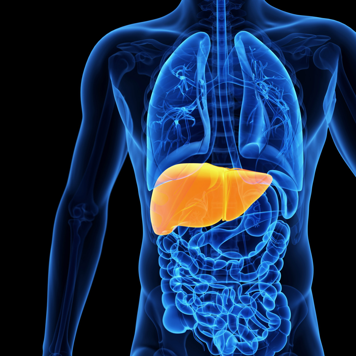
Cirrhosis, which refers to late-stage of fibrosis of the liver, occurs in approximately 5–7% of people with CF and is the third-leading cause of death in these patients. There is currently no therapy that can effectively prevent or reverse cystic fibrosis liver disease (CFLD).
Early identification of children at risk for late-stage CFLD would allow doctors to initiate preventative therapies. There is no gold-standard diagnostic test for CF-associated liver diseases. Even a liver biopsy is not diagnostically accurate for detecting CFLD.
Ultrasonography is a technique that uses echoes of ultrasound pulses to identify areas of different density. It is frequently used in the clinic and in standard monitoring of children with CF.
This imaging technique is able to detect abnormal liver tissue patterns in approximately 25% of CF patients. Still, only a minority of these patients end up developing severe liver disease.
“It is therefore imperative that we identify biomarkers that accurately reflect changes in the severity of CFLD over time, and identify patients who will progress to cirrhosis [while] they are still at an early stage of disease that is likely to be more responsive to therapeutic interventions,” researchers stated.
In the absence of a standardized method to diagnose CFLD, researchers decided to evaluate the potential of ultrasonography.
Supported by the Cystic Fibrosis Foundation and the National Institute of Diabetes and Digestive and Kidney Diseases, researchers conducted a study to investigate the clinical relevance of liver abnormalities identified by liver ultrasonography.
Specifically, researchers studied whether standard bloodwork, imaging variables, and other indicators of liver fibrosis correlated with patterns of ultrasonography in the liver. The study, named PUSH (NCT01144507), enrolled 244 children with CF, ages 3–12.
Ultrasonography findings were classified according to liver tissue pattern as either normal (NL), heterogeneous (HTG), homogeneously hyperechoic (HMG), or nodular (NOD). HTG, HMG, and NOD are all abnormal liver findings, with NOD being the most severe.
Ultrasound evaluation revealed that 122 children had normal, healthy livers, while 62 had HTG lesions, 38 had HMG, and 22 had the NOD pattern.
Results from analysis of baseline data show that there is a strong association between ultrasonography patterns and biomarkers that correlate with more advanced liver disease.
Specifically, researchers found that several biomarkers associated with more advanced liver diseases, such as aspartate aminotransferase to platelet ratio index (APRI), Fibrosis-4 score (FIB-4), and spleen size variables, correlated with patterns seen by liver ultrasonography in these patients. Blood values of AST and ALT liver enzymes, which are commonly altered when liver function is impaired, could also be linked to the different ultrasound patterns detected.
Additionally, multivariable statistical analysis of the data was able to tell apart patients with NOD from those with healthy livers with 96% accuracy. Using ultrasonography and bloodwork, the team was also able to distinguish patients with HTG and HMG from those with normal livers, and patients with NOD patterns from those with HTG, with accuracies ranging from 76–79%.
“In the absence of a gold-standard diagnostic test for CFLD and with concerns about the limited accuracy of liver biopsy for this condition, we believe that our findings validate the likely clinical significance for HTG pattern on [ultrasonography],” researchers said.
“Follow-up of this patient cohort will enable analysis of these biomarkers among children who transition between [ultrasonography] patterns over time, such as from NL to HTG and HTG to NOD,” they added.


Leave a comment
Fill in the required fields to post. Your email address will not be published.