
Computed Tomography Imaging Technique Provides Useful Insight into CF Pulmonary Damage
Written by |


According to the study “The Role of Computed Tomography in Monitoring Patients with Cystic Fibrosis” published in the Polish Journal of Radiology, computed tomography (CT), an imaging technique that is commonly used for the evaluation of cystic fibrosis (CF) in individual patients, can play a critical role in detecting lung complications and in monitoring disease progression.
CF is a genetic disorder caused by mutations in the cystic fibrosis transmembrane conductance regulator (CFTR) gene, which codes for a chloride ion channel that regulates the components of respiratory, digestive, and reproductive system secretions. When this channel is not working properly, the secretions become more viscous and the mucus builds up, particularly affecting the respiratory system by leading to chronic inflammation, bacterial colonization, and impaired airway clearance.
Over the last decades, improvement in imaging techniques, among other factors, have induced large progress in CF diagnosis and treatment strategies. In the study, Anna Rybacka and Katarzyna Karmelita-Katulska, at the Poznań University of Medical Sciences, Poland, aimed at presenting CT’s role in estimating and monitoring the course of CF lung disease severity, and assessing treatment effects.
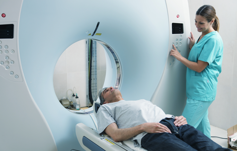
In clinical practice, the pulmonary state of patients with CF is evaluated by monitoring lung function and assessing lung structure in imaging studies. CT imaging was first used in CF patients in the 1980s, and has now become the gold standard for identification of lung structural changes in CF. It can identify bronchiectasis, the most relevant structural change in CF, characterized by permanent enlargement of parts of the airways of the lung, peribronchial thickening, mucus plugging, emphysema, and air trapping, among other disorders that occur in the progress of the disease.
Some studies have shown that CT findings were more sensitive than lung function tests. In fact, in a particular study, 48 children who were assessed by CT and pulmonary function tests at the beginning of the study and two years later, revealed progression of lung damage in the CT, whereas lung function parameters, assessed by spirometry and body plethysmography, a measurement of changes in the body’s volume, remained mostly stable.
“Because of limitations of PFT [pulmonary function tests], CT has been used more often recently to closely monitor therapeutic effects. Nowadays, CT has a crucial role in monitoring morphological changes and detecting lung complications in patients with cystic fibrosis,” Drs. Rybacka and Karmelita-Katulska concluded in their study.


Leave a comment
Fill in the required fields to post. Your email address will not be published.