Cystic Fibrosis, COPD Patients Could Benefit From Nanoparticle-based Mucus Viscosity Measuring Method


An Optical Society release notes that while people might typically regard mucus as an “icky bodily secretion best left wrapped in a tissue,” for a group of researchers from the University of North Carolina at Chapel Hill, “snot is an endlessly fascinating subject.” The team has developed a way to use gold nanoparticles and lasers to measure the viscosity (“stickiness”) of the slimy secretion that lines our airways. The new method could help doctors better monitor and treat lung diseases such as cystic fibrosis and chronic obstructive pulmonary disease.
The research team presented their work last week at The Optical Society’s 98th Annual Meeting, Frontiers in Optics, held Oct. 19-23 in Tucson, Arizona. Collocated with the American Physical Society Division of Laser Sciences 30th annual meeting, FiO brought together attendees from around the world and had more than 600 presentations covering the latest advances in all areas of optics and photonics from adaptive optics and optical sensing to silicon photonics and quantum information science. The UNC researchers’ presentation FTu5F.2, “Imaging Gold Nanorod Diffusion in Mucus Using Polarization Sensitive OCT,” was delivered Tuesday, Oct. 21 in the Tucson Ballroom, Salon A at the J.W. Marriott Tucson Starr Pass Resort.
 “People who are suffering from certain lung diseases have thickened mucus,” explains Dr. Amy Oldenburg, a physicist at the University of North Carolina at Chapel Hill whose research focuses on biomedical imaging systems. “In healthy adults, hair-like cell appendages called cilia line the airways and pull mucus out of the lungs and into the throat. But if the mucus is too viscous it can become trapped in the lungs, making breathing more difficult and also failing to remove pathogens that can cause chronic infections.”
Doctors can prescribe mucus-thinning drugs, but have up to now had no good way to monitor how they affect the viscosity of mucus at various spots inside the body. This is where Dr. Oldenburg and her colleagues’ investigations may help.
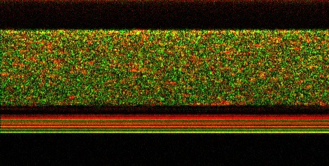
According to the Optical Society release, the researchers placed coated gold nanorods on the surface of mucus samples; then tracked the rods’ diffusion into the mucus by illuminating the samples with laser light and analyzing the way the light bounced off the nanoparticles. The slower the nanorods diffused, the thicker the mucus. The team found this imaging method worked even when mucus was sliding over a layer of cells — an important finding since mucus inside the human body is usually in motion.
“The ability to monitor how well mucus-thinning treatments are working in real-time may allow us to determine better treatments and tailor them for the individual,” says Dr. Oldenburg.
[adrotate group=”1″]
It will likely take 5 to 10 more years before the team’s mucus measuring method is tested on human patients, Dr. Oldenburg cautions. Gold is non-toxic, but for safety reasons the researchers would want to ensure that the gold nanorods would eventually be cleared from a patient’s system.
 Prof. Oldenburg is co-mentoring a project at UNC entitled “Biomedical Optics: Analyzing Dynamic Imaging Sequences of Airway Mucus Transport” with Postdoctoral Research Associate Richard Blackmon PhD. She notes that we inhale thousands of pathogens every day, which are are trapped by a mucus layer that lines the surface of our airways and acts as a protective layer.
Studying the properties of airway mucus allows scientists to understand and monitor progression of diseases, such as Chronic Obstructive Pulmonary Disease (COPD) and cystic fibrosis, that affect the respiratory system. The Oldenburg lab has been measuring mucus properties by introducing gold nanoparticles and performing optical coherence tomography (OCT) imaging.
“We have been collecting OCT images of in vitro models of the airway where mucus, bronchial cells, and nanoparticles are all moving dynamically,” says Dr. Oldenberg. “These image ‘stacks’, comprised of spatial data over time, can reveal how each of these components is moving with a different characteristic time and light signature. In this project, the student will perform analysis of dynamic OCT images (in both space and time) to look for correlations between motions of the varying components. In particular, is there any relevance to the thickness of the mucus upon the rate at which is it moving? Are the nanoparticles more or less diffusive near the mucus surface?”
 “This is a great example of interdisciplinary work in which optical scientists can meet a specific need in the clinic,” comments Nozomi Nishimura of Cornell University — one of the FiO subcommittee chairs. “As these types of optical technologies continue to make their way into medical practice, it will both expand the market for the technology as well as improve patient care.”
The Schaeffer Nishimura lab at Cornell is interested in studying the contribution of multiple physiological systems to disease initiation and progression, with applications in neurodegenerative disease, cardiovascular disease, and cancer. The team is developing delivery methods for the gold nanorods, studying how their imaging system might be adapted to enter a patients airways, and further investigating how mucus flow properties differ throughout the body. They are especially focused on understanding how the vascular, immune, inflammatory systems and cells native to a tissue interact in these diseases. The lab has developed technologies such as improved imaging using multiphoton microscopy that work in whole animals and have sufficient spatial and temporal resolution to quantify cellular dynamics. The goal of their work is to employ advanced tools like femtosecond laser ablation, to help unravel the interaction of various physiological systems in diseases, with special interests in Alzheimer’s disease, cancer metastasis, and cardiac blood flow.
Frontiers in Optics 2015 is scheduled for next October 18-22 in San Jose, California.
For more information, visit:
https://www.frontiersinoptics.com/home/
Sources:
The Optical Society
University of North Carolina at Chapel Hill
Cornell University
Image Credits:
The Optical Society
University of North Carolina at Chapel Hill
Cornell University