Cholesterol Metabolism Impaired in CF Mouse Model
Written by |

The metabolism of cholesterol is impaired in a mouse model of cystic fibrosis, a preliminary study suggests.
In the model, there seems to be an altered production of bile salts (those that help with the digestion of fats) that may reduce the digestion and/or absorption of cholesterol. This increases cholesterol’s synthesis by the liver and leads to its accumulation within cells, triggering inflammation and feeding the cycle of altered bile salt production.
The study, “Impaired cholesterol metabolism in the mouse model of cystic fibrosis. A preliminary study,” was published in the PLOS One.
CF is a is a chronic inherited disease caused by mutations in the CFTR gene. These mutations lead to the buildup of a thick mucus that changes digestion and the absorption of fatty molecules (lipids) such as cholesterol.
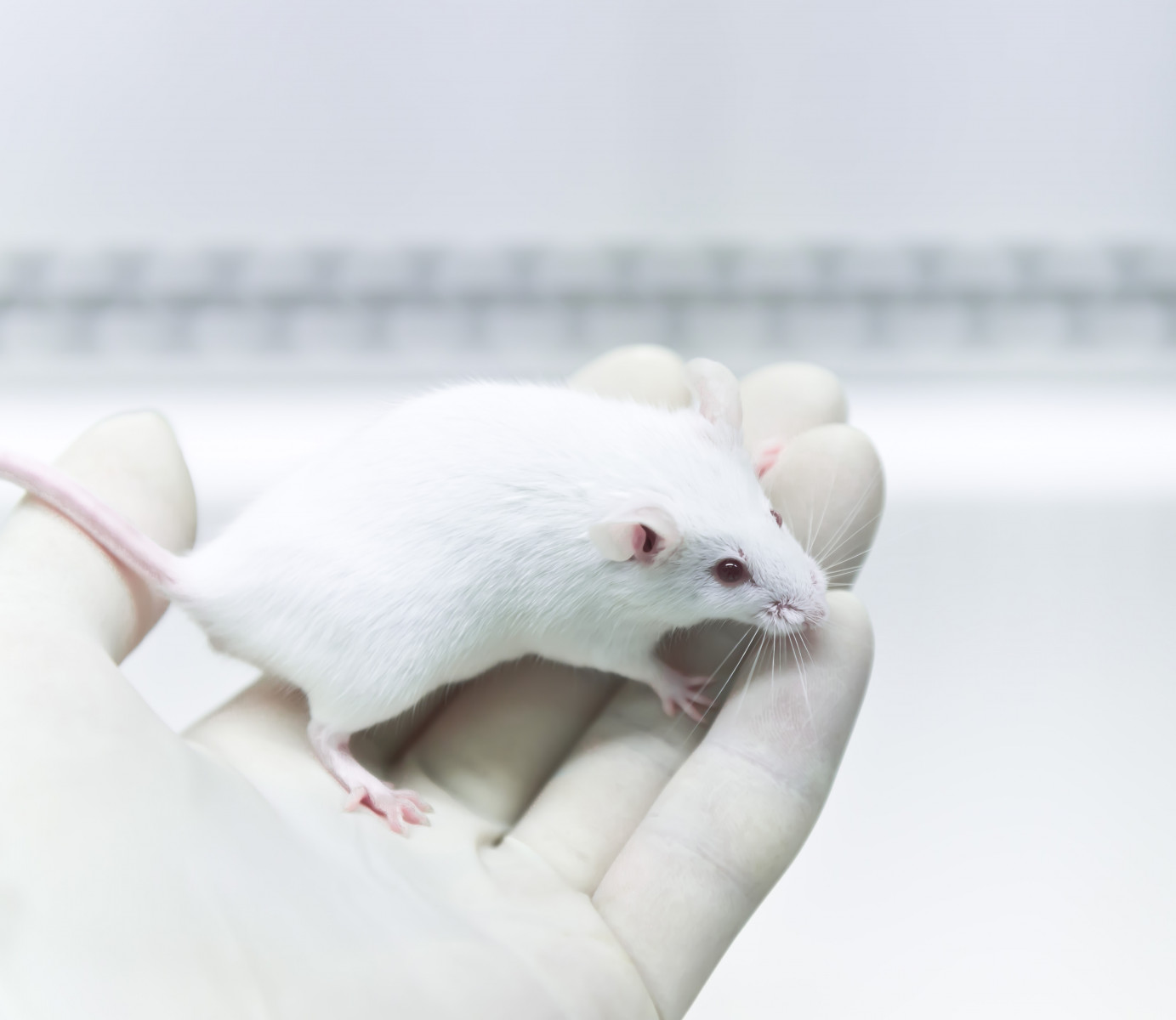
To shed light on this matter, researchers in Italy investigated cholesterol metabolism in a CF mouse model and compared it with unaffected animals. The team evaluated the effects of a diet enriched in cholesterol in both groups, and compared it with a normal diet.
Researchers analyzed blood and liver levels of several fatty molecules, including cholesterol and its derivatives phytosterols (plant-based relatives of cholesterol), lathosterol (precursor of cholesterol biosynthesis) and cholestanol (intermediate product of bile salts synthesis).
The team also measured the expression of different genes, including LDLR, HMG-CoAR, ACAT2, CYP7A1 and TNFA, in the liver. These genes provide instructions for the production of key molecules involved in the metabolism of cholesterol. Gene expression is the process by which information in a gene is synthesized to create a working product, like a protein. Tumor necrosis factor alpha (TNF-alpha), the product of TNFA, is an inflammation marker.
Mice were fed either with a diet enriched with linoleic acid, a fatty molecule, and vitamin E for 14 weeks (three mice with CF and three unaffected) or for 10 weeks. After that they were fed a diet enriched in cholesterol for four weeks (four mice with CF and four unaffected).
Compared to those fed without cholesterol supplementation, cholesterol-supplemented healthy mice showed significantly lower levels of blood phytosterols (7.6 times lower) and liver lathosterol (7.5 times), and higher levels of liver cholesterol (6.2 times higher) and cholestanol (4.2 times). Blood cholesterol was significantly higher (1.3 times) in supplemented mice.
Furthermore, unaffected mice fed with cholesterol supplementation had significantly lower expression levels of LDLR (2.5 times) and HMG-CoAR (2.2 times), while ACAT2 and CYP7A1 expression levels were significantly higher (2.9 and 6.0 times, respectively). The expression levels of TNFA showed an increasing trend in cholesterol-supplemented unaffected mice.
CF mice that did not receive cholesterol supplementation had significantly lower blood levels of phytosterols (2.0 times) and a decreasing trend of blood cholesterol when compared to healthy mice fed the same diet. In contrast, lathosterol and cholesterol levels in the liver were significantly higher (3.1 and 1.2 times, respectively) with an increasing trend of liver cholestanol.
CF mice had significantly higher gene expression levels of HMG-CoAR (18.0 times) and CYP7A1 (16.3 times) with an increasing trend of LDLR and ACAT2 gene expression levels in comparison with unaffected mice. Expression levels of TNFA showed an increasing trend in CF mice.
The team then evaluated the effects of cholesterol supplementation in CF mice. When compared with healthy mice that also received supplementation, CF mice showed significantly higher blood levels of phytosterols and liver lathosterol (2.0 and 22.0 times, respectively), significantly lower levels of blood and liver cholesterol (1.3 and 4.3 times, respectively) and significantly lower levels of liver cholestanol (3.4 times).
Additionally, the supplemented CF mice had significantly higher gene expression levels of LDLR (4.7 times), HMG-CoAR (41.2 times) and CYP7A1 (5.3 times) when compared with supplemented unaffected mice, while ACAT2 expression levels were not significantly different. Expression levels of TNFA were higher in CF mice, but not significant.
When comparing the CF mice with and without supplementation, there were no significant differences in the parameters studied, except for the levels of blood phytosterols that were significantly lower in the supplemented CF mice, and liver cholesterol and TNF-alpha levels that were significantly higher in the supplemented CF mice.
“Our results show that in CF mice there is an impairment of intestinal cholesterol absorption and liver cholesterol metabolism,” the researchers wrote.
Although these results should be confirmed by future experiments, the team believes that “this preliminary study suggests that in CF mice there is a vicious circle in which the altered synthesis/secretion of bile salts may reduce the digestion/absorption of cholesterol. As a result, the liver increases the biosynthesis of cholesterol that accumulates in the cells, triggering inflammation and further compromising the metabolism of bile salts.”


