
Microsoft’s Xbox Kinect Devices Used to Develop New Way of Assessing CF Lung Health
Written by |
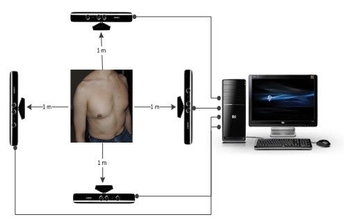
Kinect technology being used to assess respiratory patients with CF and other lung conditions. (Image credit: University of Warwick)


A new technique using relatively inexpensive Microsoft Xbox Kinect gaming devices could soon be used to assess the respiratory health of patients with conditions such as cystic fibrosis (CF).
Researchers at the Institute of Digital Healthcare, WMG at the University of Warwick in Coventry, England, the Institute of Inflammation and Aging at the University of Birmingham, and the Heart of England NHS Foundation Trust (HEFT) have developed a method of using Kinect technology to assess respiratory patients with CF and other lung conditions. The experimental system consists of four Kinect sensors that are capable of quickly creating 3-D imaging of a patient’s torso, enabling physicians to measure and assess the subject’s chest wall motion.
In tests, the technique has proven to be as accurate as having a patient breath into a spirometer — the current standard method for assessing lung diseases such as chronic bronchitis, emphysema, and chronic obstructive airways — as well as providing additional information about chest movement that could help in identifying a range of respiratory problems.

Image Credit: University of Warwick
Spirometry requires patients to inhale the deepest breath they can, then exhale into the sensor as vigorously as possible, for as long as possible. However, the researchers note that spirometry has significant limitations, particularly in not enabling doctors to assess how different areas of each lung are functioning. As a result, it can yield inaccurate readings for some patients, particularly older people and children, as well as people with facial abnormalities or muscle weakness that render them unable to form a tight seal around the spirometer’s mouthpiece.
Referring to the experimental Kinect-based system, the project’s leader, Dr. Chris Golby of the Institute of Digital Healthcare, said: “We have developed a low-cost prototype which provides a more comprehensive measurement of a patient’s breathing then existing methods.”
The research team’s work is detailed in an Open Access paper titled “Chest wall motion analysis in healthy volunteers and adults with cystic fibrosis using a novel Kinect-based motion tracking system” (DOI: 10.11007/s11517-015-1433-1), and published in the journal Medical & Biological Engineering & Computing. The paper was co-authored by Dr. Golby, James M. Harte and Theodoros N. Arvanitis of the Institute of Digital Healthcare, WMG, University of Warwick Interacoustics Research Unit, c/o Technical University of Denmark; Johanna Acosta of the Institute of Digital Healthcare, WMG, University of Warwick; Edward F Nash of the Heart of England NHS Foundation Trust; Ercihan Kiraci and Mark A. Williams of WMG, University of Warwick; and Babu Naidu of the Heart of England NHS Foundation Trust at the University of Birmingham.
The researchers note that respiratory disease is the leading cause of death in the U.K., accounting for 920,000 disability-adjusted life years lost annually — and is the most frequent cause of disease in primary care in all age groups, and the second most common cause of chronic conditions. Methods for assessing pulmonary function and chest wall movement are essential for accurate diagnosis, as well as for monitoring response to treatment, operative procedures, and rehabilitation. Despite this need, they observe that there is a lack of low-cost devices for rapid lung assessment, and spirometry is unable to directly measure full chest wall motion.
The study details the development of the low-cost chest wall motion assessment system using the four Microsoft Kinect sensors, as well as evaluation of the system in two phases. The co-authors report that initially, static volume of a resuscitation mannequin was performed with a Nikon laser scanner, which showed that the system has a slight under-prediction of 0.441 percent. Next, a dynamic analysis was performed comparing results from the prototype Kinect-based system and from a spirometer in nine cystic fibrosis patients and 13 healthy subjects. The comparison showed agreement of the two methods, with correlation coefficients above 0.8656 in all participants.
“For patients … a quick and low-cost method of chest wall motion assessment is required,” says Dr. Golby in a University of Warwick release. “There are some conditions that doctors can’t detect or assess using spirometry such as collapsed lung segments or respiratory muscle weakness. However our prototype allows physicians to make accurate assessments. It is also potentially very useful in assessing changes in respiratory physiology that occur during exercise. This is in contrast with existing systems which rely on data from one viewpoint.”
 Paper co-author and chief investigator Babu Naidu, a thoracic surgeon at HEFT and clinical scientist at the University Birmingham, calls the Kinect-based system “a ‘game changer’ in screening, diagnostics, monitoring therapy and providing bio feedback,” observing that “the Xbox can be used in any condition affecting breathing.”
The researchers note that while respiratory diseases kill 1 in 5 people in the U.K. and cost the National Health Service (NHS) more than £6 billion a year, the proposed system’s software and four Kinect sensors would each cost just £100.
 Professor Theo Arvanitis, another paper co-author and head of research at the WMG Institute of Digital Healthcare, observes, “With this and other technologies developed, we hope to innovate in e-healthcare and translate these advances into clinical practice.”
The investigators, in their study on a resuscitation mannequin and in human participants, reported that the Kinect’s infrared beam enabled them to measure changes in distances across the chest wall, since its four sensors allow measurement of movement from more than one perspective. Using off-the-shelf and 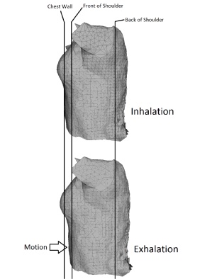 bespoke software, the researchers were able to create 3-D images of patient’s chest walls. They noted that another advantage of using data from four Kinect sensors is that this system is able to accurately assess chest wall motion even in moving subjects, a capability potentially very useful for assessing changes in respiratory physiology that occur on exercise, such as dynamic hyperinflation. (Image Credit: University of Warwick)
Consequently, the investigators concluded that these initial results suggest that the Kinect-based system is an accurate and affordable alternate method of rapidly measuring chest wall motion, although more work must be done to develop and evaluate the prototype. The University of Warwick team is now planning to develop their prototype further using Microsoft’s latest version of the Kinect device and SDK, and to evaluate the system for testing regional motion and tele-assessments. The team will also be evaluating it in comparison with optoelectronic plethysmography, and there will be further tests involving people with CF and other respiratory conditions.
Sources:
University of Warwick
University of Birmingham
Institute of Digital Healthcare, WMG
Heart of England NHS Foundation Trust


Leave a comment
Fill in the required fields to post. Your email address will not be published.